
新聞與活動
探索尚博生物科技的最新動態、技術新知與產業活動
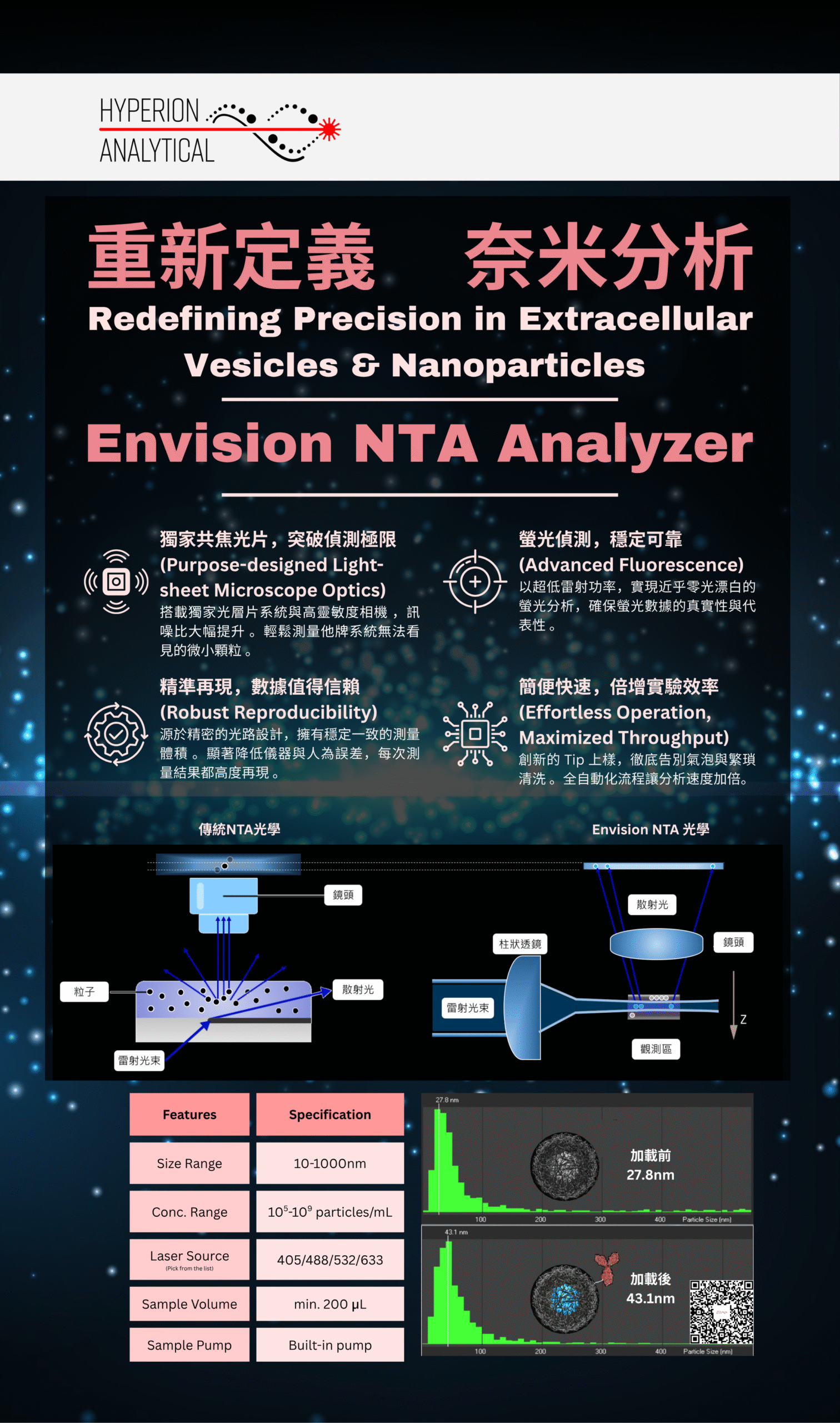
重新定義 奈米分析:Envision NTA Analyzer
次世代光層片NTA 分析儀,透過獨特的光學設計,大幅提升奈米顆粒分析的精準度與敏感度;Build-in pump 避免手動注入樣本所造成的誤差...
閱讀更多 →
探索尚博生物科技的最新動態、技術新知與產業活動
次世代光層片NTA 分析儀,透過獨特的光學設計,大幅提升奈米顆粒分析的精準度與敏感度;Build-in pump 避免手動注入樣本所造成的誤差...
閱讀更多 →